Home
/ Cross Section Of A Bone Diagram : Part 2 at University of North Carolina-Pembrok - StudyBlue : The periosteum contains many strong collagen fibers that are used to firmly anchor tendons and muscles to the bone for movement.
Cross Section Of A Bone Diagram : Part 2 at University of North Carolina-Pembrok - StudyBlue : The periosteum contains many strong collagen fibers that are used to firmly anchor tendons and muscles to the bone for movement.
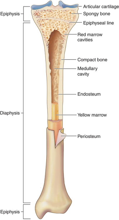
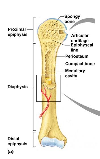
Cross Section Of A Bone Diagram : Part 2 at University of North Carolina-Pembrok - StudyBlue : The periosteum contains many strong collagen fibers that are used to firmly anchor tendons and muscles to the bone for movement.. There are two ways to study bone histology. Diagram with articular cartilage, marrow, medullary cavity and periosteum. Looking at a bone in cross section, there are several distinct layered regions that make up a bone. Contact your company to license this image. Start studying cross section of bone.
Indicated are haversian canal with blood and lymphatic vessels, a nerve, and loose. Volcano cross section diagram drawing high. A cross section of a compact bone shows concentric circles called lamellae. The maxillary bones important structures. Bone markings the surface features of bones vary considerably, depending on the function and location in the body.
Structure and Function of the Musculoskeletal System ... from basicmedicalkey.com Diagram with articular cartilage, marrow, medullary cavity and periosteum. Learn vocabulary, terms, and more with flashcards, games, and other study tools. Related posts of cross section of human bone diagram human back muscles and bones. A diagram of the anatomy of a bone, showing the compact bone. Human bone, cross section diagram of femur showing osteon, veins, marrow. Cross section of the leg through the soleus muscle: Two types of bone tissues in cross section of a long bone : (b) in this micrograph of the osteon, you can clearly see the concentric lamellae and central canals.
Indicated are haversian canal with blood and lymphatic vessels, a nerve, and loose.
Helps form the zygomatic arches. Bone cross section diagram ipad folio cases. Bone markings the surface features of bones vary considerably, depending on the function and location in the body. Start studying cross section of bone. Bone on side of the foot 12 photos of the bone cross section labeled. Cross section of a bone diagram. Learn vocabulary, terms, and more with flashcards, games, and other study tools. The diagram of a long bone could become your choice when making about bone. Foot bone anatomy x ray 12 photos of the foot bone anatomy x ray foot bone anatomy x ray, bone, foot bone anatomy x ray. Looking at a bone in cross section, there are several distinct layered regions that make up a bone. The outside of a bone is covered in a thin layer of dense irregular connective tissue called the periosteum. Related posts of cross section of human bone diagram human back muscles and bones.
The structures and function of bones, bone marrow, and cartilage; 12 photos of the bone cross section labeled. (b) in this micrograph of the osteon, you can clearly see the concentric lamellae and central canals. Cross section of a bone / long bone human skeleton fraudbein cross section png 1000x500px watercolor cartoon flower frame. Bone on side of the foot
Print Exercise 9: Overview of the Skeleton: Classification ... from www.easynotecards.com Generally speaking, it is very easy to recognize a cross section through the leg, mostly due to the tibia. Looking at a bone in cross section, there are several distinct layered regions that make up a bone. This is the same reason why the slightest touch hurts so much. Cross section = transverse section bone cross section. Indicated are haversian canal with blood and lymphatic vessels, a nerve, and loose. Related posts of cross section of human bone diagram human back muscles and bones. Learn vocabulary, terms, and more with flashcards, games, and other study tools. Helps form the zygomatic arches.
The structures and function of bones, bone marrow, and cartilage;
Start studying cross section of bone. Cross section of a bone : This bone is located directly beneath the skin on the anterior aspect of the leg (top of the image). Diagram with articular cartilage, marrow, medullary cavity and periosteum. Volcano cross section diagram drawing high. Long bones grow more than the other classes of bone throughout childhood and so are responsible for the bulk of our height as adults. Helps form the zygomatic arches. The periosteum contains many strong collagen fibers that are used to firmly anchor tendons and muscles to the bone for movement. The development aspects of bones The lower set of teeth in the mouth is rooted in the lower jaw. Download scientific diagram | schematic of osteon. (b) in this micrograph of the osteon, you can clearly see the concentric lamellae and central canals. Learn vocabulary, terms, and more with flashcards, games, and other study tools.
Bone on side of the foot For example, to read this diagram literally, since the cartilage can be seen inside the cutaway section of bone, it incorrectly indicates that the cartilage in fact goes through the bone structure, rather than just being found around the bone end. This is known as the periosteum. Indicated are haversian canal with blood and lymphatic vessels, a nerve, and loose. Browse 4,280 bone cross section stock photos and images available, or search for human bone cross section to find more great stock photos and pictures.
Ch. 12 Spinal Cord Objectives at Mount Royal University ... from classconnection.s3.amazonaws.com Start studying cross section of bone. Indicated are haversian canal with blood and lymphatic vessels, a nerve, and loose. Cross section of a bone : Related posts of cross section of a long bone foot bone anatomy x ray. The development aspects of bones This is known as the periosteum. Bodytomy provides a labeled diagram of the haversian system to help you the terms 'haversian system' or 'osteon' refer to the basic. The name, structure, and function of joint types and the ranges allowed by each joint;
Download scientific diagram | schematic of osteon.
This is known as the periosteum. An outer 'fibrous layer' containing mainly fibroblasts, and an inner 'cambium layer' containing progenitor cells. Smartdraw includes 1000s of professional healthcare and anatomy chart templates that you can modify and make your own. Human bone, cross section diagram of femur showing osteon, veins, marrow. Diagram with articular cartilage, marrow, medullary cavity and periosteum. Learn vocabulary, terms, and more with flashcards, games, and other study tools. Bodytomy provides a labeled diagram of the haversian system to help you the terms 'haversian system' or 'osteon' refer to the basic. Long bones grow more than the other classes of bone throughout childhood and so are responsible for the bulk of our height as adults. The line will be indicated by an actual line, or with. Über 7 millionen englischsprachige bücher. Bone on side of the foot (b) in this micrograph of the osteon, you can clearly see the concentric lamellae and central canals. Bone cross section diagram ipad folio cases.
Start studying cross section of long bone cross section of a bone. Bone markings the surface features of bones vary considerably, depending on the function and location in the body.